Nanosensors for biomedical applications

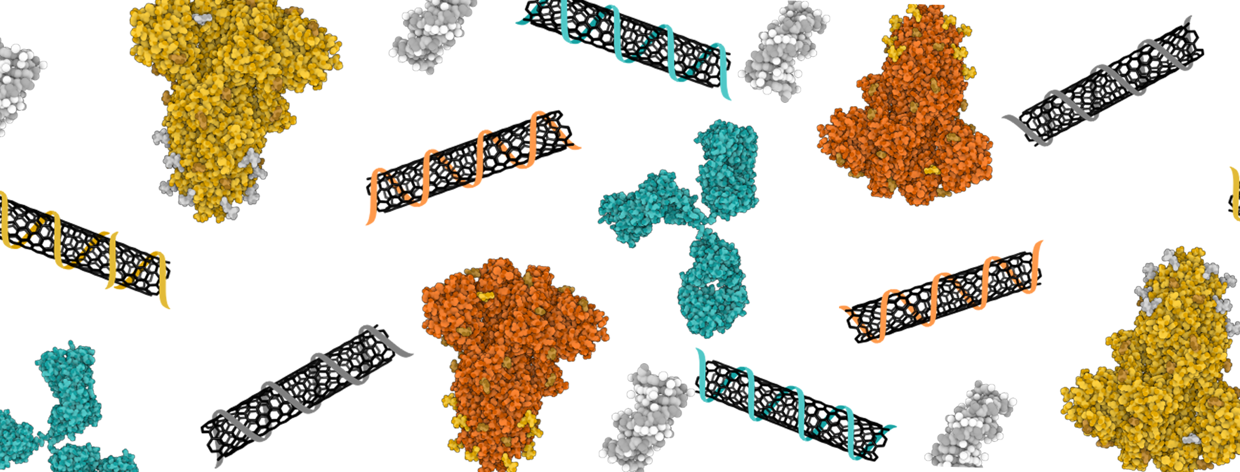
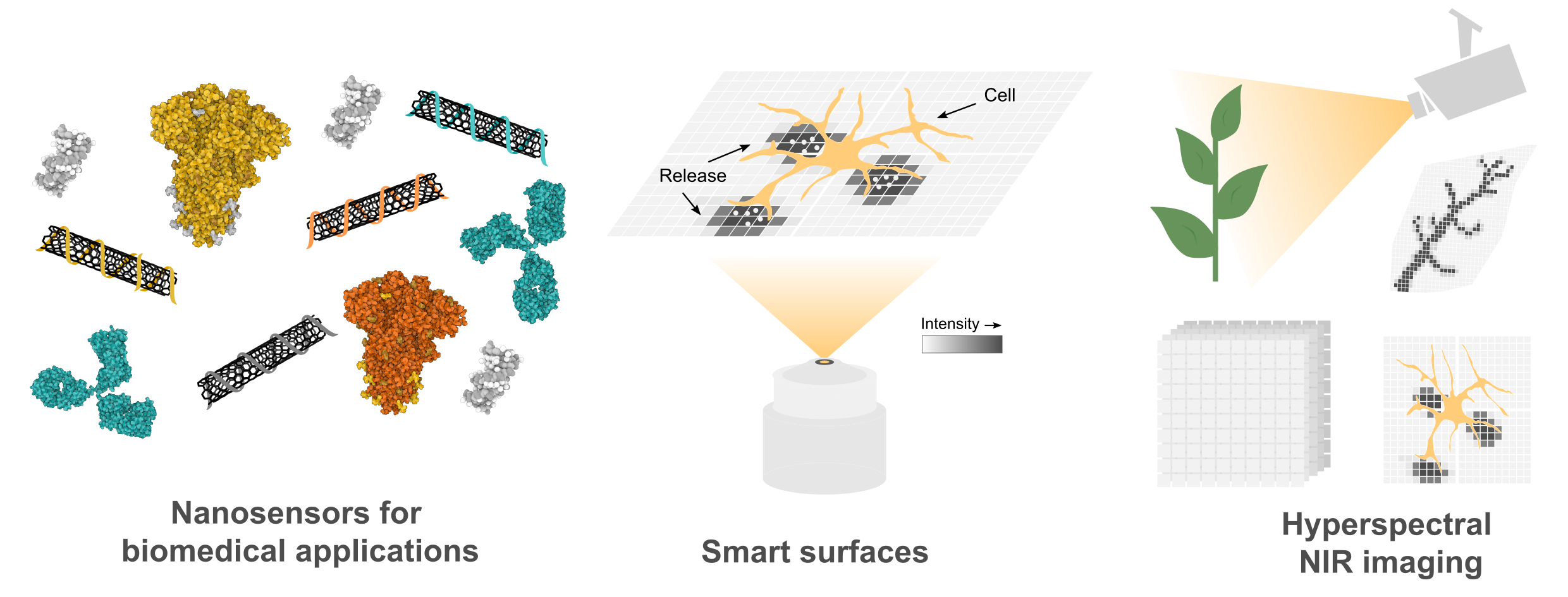
The rapid and specific detection of chemical species is one of the key technologies of the 21st century. It presents the basis for advances in molecular diagnostics, personalized medicine, and environmental monitoring. In medical diagnostics, the analysis of tissues and biological fluids such as blood or saliva plays a central role for the detection of infections or the monitoring the of diseases and treatment plans. That said, sensors for these areas are essential tools for the rapid detection of pathogens and pollutants or for process control in chemical and biotechnological processes. The urgent need for flexible, sensitive detection solutions has become particularly apparent during the SARS-CoV-2 pandemic.

At Fraunhofer IMS, we develop sensors for medical use that can provide valuable services in biological applications. One focus is on optical sensors that can measure through tissue or body fluids such as blood or saliva without direct contact. They use special fluorescence signals in the near-infrared range (780-2400 nm), which can be detected through deeper layers if tissue. This allows important information about a person's state of health to be recorded quickly and accurately and can be used for the detection of infections, the monitoring of disease progression, or to support individual therapies.

© Fraunhofer IMS
An overview of the group's research areas.

At a glance

Customer benefits

  • Fast, precise, and contact-free detection of a wide range of analytes
  • High sensitivity and flexibility
  • Integration into existing workflows
  • Customizable development of sensors and optical systems

Technologies

  • Optical sensors with near-infrared fluorescence (780–2400 nm)
  • Contactless measurement through tissue or liquids
  • Compact, integrable sensor components

Applications

  • Infection diagnostics and disease monitoring
  • Environmental and pollutant detection
  • Process control in biotechnology and chemical engineering

Our research areas

© Sebastian Kruss/RUB
© Fraunhofer IMS
Optical nanosensors with carbon nanotubes (SWCNTs). By functionalizing SWCNTs with various (bio)polymers, modular sensors can be produced for a wide range of analytes. The interaction between the analytes and the sensors leads to a change in fluorescence.
© 2025 The Author(s). Angewandte Chemie International Edition published by Wiley-VCH GmbH

At Fraunhofer IMS, we develop biofunctional sensors for applications in medical diagnostics. We use semiconducting carbon nanotubes (single-walled carbon nanotubes, SWCNTs) that fluoresce in the near infrared (NIR, 870-2400 nm). Unlike other fluorophores, they do not bleach, which makes them ideal for long-term monitoring.

Our sensor technology is based on changes in the molecular environment of one-dimensional nanomaterials. The sensors can be assembled in a modular fashion.

In addition, the surface of the nanotubes is highly sensitive to changes in their environment. Through clever design, sensors can be created for a wide range of analytes. Our applications range from the spatially and temporally resolved detection of neurotransmitter release and the detection of the SARS-CoV-2 spike protein to the detection of bacteria in culture supernatants and the monitoring of enzymatic reactions.

We are constantly working on innovative approaches to improve the performance of our sensors. This includes modifying the sensors with so-called quantum defects, which can be imagined as small irregularities in an otherwise perfectly structured material.

By introducing such irregularities in a controlled manner, the optical properties can be controlled and optimized for various applications. For example, it is possible to modify the fluorescence of the nanotubes by changing their absorption and emission wavelengths and to increase the quantum efficiency, i.e., the intensity of the fluorescence.

The introduction of defects furthermore allows us to functionalize nanosensors with detection units and to control the selectivity of one-dimensional nanosensors by structuring the surface at the molecular level.

Here you can learn how visible light limits applications in the biomedical field and how you can use signals in the NIR to see deeper into tissue and biological fluids.

© Justus Metternich/Fraunhofer IMS
Photo of a red wine bottle, a red wine glass, and a cactus in the visible (top) and NIR range (bottom). Similar to wine, tissues are more permeable in the NIR range, enabling our sensors to read through tissues and liquids without contact.
© Justus Metternich/Fraunhofer IMS

In modern sensor technology, choosing the optimal wavelength for detection plays a crucial role. Due to the optical properties of biological samples, selecting the optimal wavelength is particularly important in the biomedical field.

While visible light is widely used in many applications, it often reaches its limits when it comes to examining biological tissues and fluids.

NIR sensors enable deeper tissue layers to be reached, as absorption, scattering, and autofluorescence are significantly reduced compared to the visible spectrum. Optical sensors that fluoresce in the NIR therefore offer decisive advantages for contactless readout in biomedical applications.

At Fraunhofer IMS, we develop sensor technology specialized for biomedical applications in the NIR range. Thanks to their platform character, our sensors can be adapted quickly and cost-effectively. Please do not hesitate to contact us if you have any questions about the development of further sensors.

Don't have an optical readout unit for NIR yet? In addition to our sensors, we also develop optical detection systems specifically for the NIR range.

© 2025 The Authors. Advanced Science published by Wiley-VCH GmbH (CC BY 4.0). Adapted.
HyperNIR can be used to visualize how a pepper plants absorb water. On the right are three images of a single leaf and the corresponding HyperNIR images, which visualize water absorption; the redder the color, the more water is present at that point on the leaf.

The optical detection of signals in the near-infrared (NIR) spectral range (780-2400 nm) enables particularly interference-free readouts through biological tissue and fluids in biomedical contexts, characterized by high signal-to-noise ratios and deep penetration depths.

In order to detect and image the signals from optical sensors, we at Fraunhofer IMS develop innovative setups that enable you to read signals in the NIR range.

These systems include compact readout devices for the spectral range > 900 nm and hyperspectral setups that can generate rapid hyperspectral (NIR) images with only three images, as well as the modification of standard microscopes for the NIR range.

Hyperspectral imaging with three images

Hyperspectral imaging captures both spectral and spatial information. Our HyperNIR module requires only three images and generates spectral information in the range between 900 nm and 1600 nm. Spectral resolutions of less than 5 nm and frame rates of 0.2 frames per second are achieved using the full camera resolution.

This user-friendly technology enables simple hyperspectral imaging, whereby any standard camera for the NIR range can be upgraded to a hyperspectral camera using the HyperNIR module. This allows us to access wide range of biomedical imaging and environmental monitoring applications.

Highlights from our research


Smart Slides – biomolecular NIR platform for cell culture

Nanosensors in surface coatings enable molecular imaging of release processes with high spatial and temporal resolution.


Sensor-based signal amplification of biological assays

Our nanosensors react to enzymatic substrates and products used in diagnostics. Upon simple addition of the sensors signals of a reaction can be amplified by up to 120-fold. Adding our sensors during the reaction allows you to track enzymatic processes below the classic detection limit.


Sensors for detecting pathogens and monitoring inflammatory processes

By combining several nanosensors, we are able to distinguish between different bacteria. We are currently working on solutions for inflammatory processes and contamination monitoring in cell cultures.


Purified NIR sensors for the detection of neurotransmitters

We offer purified (6,4)- and (6,5)-carbon nanotubes whose fluorescence lies in the transition range between NIR and VIS. This allows the advantages of the NIR range to be combined with the use of standard laboratory equipment. On request, purification processes for other nanotubes, such as metallic chiralities, can also be developed.


HyperNIR: Hyperspectral imaging in the tissue transparency window

Near-infrared light, which is invisible to humans, contains valuable information about the chemical composition of samples. With the real-time method we have developed, microplastics, plant stress, or pathogens can be detected in environmental monitoring, for example, and the chemical composition of various samples can be determined. The system can be adapted for different cameras on request.

Scientific publications

Here you can find further current scientific publications:

Jahr
Year
Titel/Autor:in
Title/Author
Publikationstyp
Publication Type
2025 3D printing of models of carbon nanotubes and related nanomaterials
Gretz, Juliana; Kruss, Sebastian
Zeitschriftenaufsatz
Journal Article
2025 High-Speed Hyperspectral Imaging for Near Infrared Fluorescence and Environmental Monitoring
Stegemann, Jan; Gröniger, Franziska; Neutsch, Krisztian; Li, Han; Flavel, Benjamin Scott; Metternich, Justus Tom; Erpenbeck, Luise; Petersen, Poul Bering; Hedde, Per Niklas; Kruss, Sebastian
Zeitschriftenaufsatz
Journal Article
2025 Solvatochromic Dyes Increase the Sensitivity of Nanosensors
Ma, Chen; Kistwal, Tanuja; Hill, Björn F.; Neutsch, Krisztian; Kruss, Sebastian
Zeitschriftenaufsatz
Journal Article
2025 Levodopa Sensing with a Nanosensor Array via a Low-Cost Near Infrared Readout
Stegemann, Jan; Augustin, Matthias Niklas; Ackermann, Julia; Fizzi, Nour El Houda; Neutsch, Krisztian; Gregor, Markus; Herbertz, Svenja; Kruss, Sebastian
Zeitschriftenaufsatz
Journal Article
2024 Fluorescence changes in carbon nanotube sensors correlate with THz absorption of hydration
Nalige, Sanjana S.; Galonska, Phillip; Kelich, Payam; Sistemich, Linda; Herrmann, Christian; Vukovic, Lela; Kruss, Sebastian; Havenith, Martina
Zeitschriftenaufsatz
Journal Article
2024 Hyperspectral near infrared imaging using a tunable spectral phasor
Stegemann, Jan; Gröniger, Franziska; Neutsch, Krisztian; Li, Han; Flavel, Benjamin; Metternich, Justus Tom; Erpenbeck, Luise; Petersen, Poul; Hedde, Per Niklas; Kruss, Sebastian
Paper
2024 High Throughput Approaches to Engineer Fluorescent Nanosensors
Metternich, Justus Tom; Patjoshi, Sujit K.; Kistwal, Tanuja; Kruss, Sebastian
Paper
2024 Verfahren zum Funktionalisieren eines Kohlenstoffnanoröhrchen-Materials, funktionalisiertes CNT-Material und Sensoren mit einem CNT-Material
Kruss, Sebastian; Metternich, Justus Tom; Herbertz, Svenja
Patent
2024 Size Matters in Conjugated Polymer Chirality-Selective SWCNT Extraction
Dzienia, Andrzej; Just, Dominik; Wasiak, Tomasz; Milowska, Karolina Z.; Mielańczyk, Anna; Labedzki, Norman; Kruss, Sebastian; Janas, Dawid
Zeitschriftenaufsatz
Journal Article
2024 Ratiometric Near Infrared Fluorescence Imaging of Dopamine with 1D and 2D nanomaterials
Hill, Bjoern Fabian; Mohr, Jennifer Maria; Sandvoß, Isabelle K.; Gretz, Juliana; Galonska, Phillip; Schnitzler, Lena; Erpenbeck, Luise; Kruss, Sebastian
Paper
Diese Liste ist ein Auszug aus der Publikationsplattform Fraunhofer-Publica

This list has been generated from the publication platform Fraunhofer-Publica


Learn more

You can find more information about our university group and our research here:


Technology (Home)

This will take you back to the overview page for the core competency Technology.

Kontakt

Prof. Anna Lena Schall-Giesecke

Core Competence Technology

Fraunhofer-Institute for Microelectronic Circuits and Systems IMS
Finkenstraße 61
47057 Duisburg, Germany